Modern and Innovative Foot, Ankle, and Leg Treatments
Since 2001, Southeast Foot & Ankle has been a preferred treatment center for a wide range of conditions that cause pain and lack of mobility. We have always provided scientifically backed treatments, effective sports medicine, as well as other solutions to various ailments and injuries.
On top of our knowledge and resources, another factor that sets us apart from other treatment facilities is our unwavering commitment to our patients. We truly care about your health and comfort, which is why we constantly strive to educate ourselves on the latest foot, ankle, and leg treatments.
If you come to us, you will be in the hands of a skilled, dedicated, and well-rounded team. Here are just several of the conditions that we treat:
- Custom Orthotics
- Diabetic Foot Care
- Sports injuries
- Sports Medicine and Related Injury
- Tendinitis
- Wart Removal
- Wound Care
We serve patients based in several locations in Alaska, particularly Ketchikan, Sitka, and Juneau. Reach out to us today!

Accessory Navicular Syndrome
What is the Accessorv Navicular?
The accessory navicular (os navicularum or os tibiale exEmum) is an extra bone or piece of cartilage located on the inner side of the foot just above the arch. It is incorporated within the posterior tibial tendon, which attaches in this area. An accessory navicular is congenital (present at birth), It is not part of normal bone structure and therefore is not present in most people
What is Accessory Navicular Svndrome?
People who have an accessory navicular often are unaware of the condition if it causes no problems.
However, some people with this extra bone develop a painful condition known as accessory navicular syndrome when the bone and/or posterior tibial tendon are aggravated. This can result from any of the following:
- Trauma, as in a foot or ankle sprain
- Chronic irritation from shoes or other footwear rubbing against the extra bone
- Excessive activity or overuse
Many people with accessory navicular syndrome also have flat feet (fallen arches). Having a flat foot puts more strain on the posterior tibial tendon, which can produce inflammation or irritation of the accessory navicular.
Signs and Symptoms of Accessory Navicular Syndrome
Adolescence is a common time for the symptoms to first appear. This is a time when bones are maturing and cartilage is developing into bone. Sometimes, however, the symptoms do not occur until adulthood. The signs and symptoms of accessory navicular syndrome include:
- A visible bony prominence on the midfoot (the inner side of the foot, just above the arch)
- Redness and swelling of the bony prominence
- Vague pain or throbbing in the midfoot and arch, usually occurring during or after periods of activity
Diagnosis
To diagnose accessory navicular syndrome, the foot and ankle surgeon will ask about symptoms and examine the foot, looking for skin irritation or swelling. The doctor may press on the bony prominence to assess the area for discomfort. Foot structure, muscle strength, joint motion, and the way the patient walks may also be evaluated.
X—rays are usually ordered to confirm the diagnosis. If there is ongoing pain or inflammation, an MRI or other advanced imaging tests may be used to further evaluate the condition.
Treatment: Non-Surgical Approaches
The goal of nonsurgical treatment for accessory navicular syndrome is to relieve the symptoms. The following may be used:
- Immobilization. Placing the foot in a cast or removable walking boot allows the affected area to rest and decreases the inflammation.
- Ice. To reduce swelling, a bag of ice covered with a thin towel is applied to the affected area. Do not put ice directly on the skin.
- Medications. Oral no steroidal anti—inflammatory drugs (NSAIDs), such as ibuprofen, may be prescribed. In some cases, oral or injected steroid medications may be used in combination with immobilization to reduce pain and inflammation.
- Physical therapy. Physical therapy may be prescribed, including exercises and treatments to strengthen the muscles and decrease inflammation. The exercises may also help prevent recurrence of the symptoms.
- Orthotic devices. Custom orthotic devices that fit into the shoe provide support for the arch, and may play a role in preventing future symptoms. Even after successful treatment, the symptoms of accessory navicular syndrome sometimes reappear. When this happens, non—surgical approaches are usually repeated.
When is Surgery Needed?
If non-surgical treatment fails to relieve the symptoms of accessory navicular syndrome, surgery may be appropriate. Surgery may involve removing the accessory bone, reshaping the area, and repairing the posterior tibial tendon to improve its function. This extra bone is not needed for normal foot function.
Achilles Tendon Rupture
What is the Achilles Tendon?
A Tendon is a band of tissue that connects a muscle to a bone. The Achilles tendon runs down the back of the lower leg and connects the calf muscle to the heel bone. Also called the "heel cord,” the Achilles tendon facilitates walking by helping to raise the heel off the ground.
What is an Achilles Tendon Rupture?
An Achilles tendon rupture is a complete or partial tear that occurs when the tendon is stretched beyond its capacity. Forceful jumping or pivoting, or sudden accelerations of running, can overstretch the tendon and cause a tear. An injury to the tendon can also result from falling or tripping.
Achilles tendon ruptures are most often seen in "weekend warriors” —— typically, middle—aged people participating in sports in their spare time; Less commonly, illness or medications, such as steroids or certain antibiotics, may weaken the tendon and contribute to ruptures.
Signs and Symptoms
A person with a ruptured Achilles tendon may experience one or more of the following:
- Sudden pain (which feels like a kick or a stab) in the back of the ankle or calf—often subsiding into a dull ache
- A popping or snapping sensation
- Swelling on the back of the leg between the heel and the calf
- Difficulty walking (especially upstairs or uphill) and difficulty rising up on the toes
These symptoms require prompt medical attention to prevent further damage. Until the patient is able to see a doctor, the”RICE.” Method should be used. This involves:
- Rest. Stay off the injured foot and ankle, since walking can cause pain or further damage.
- Ice. Apply a bag of ice covered with a thin towel to reduce swelling and pain. Do not put ice directly against the skin.
- Compression. Wrap the foot and ankle in an elastic bandage to prevent further swelling.
- Elevation. Keep the leg elevated to reduce the swelling. It should be even with or slightly above heart level.
Diagnosis
In diagnosing an Achilles tendon rupture, the foot and ankle surgeon will ask questions about how and when the injury occurred and whether the patient has previously injured the tendon or experienced similar symptoms.
The surgeon Will examine the foot and ankle, feeling for a defect in the tendon that suggests a tear. Range of motion and muscle strength will be evaluated and compared to the uninjured foot and ankle. If the Achilles tendon is ruptured, the patient will have less strength in pushing down (as on a gas pedal) and will have difficulty rising on the toes.
The diagnosis of an Achilles tendon rupture is typically straightforward and can be made through this type of examination. In some cases, however, the surgeon may order an MRI or other advanced imaging tests.
Treatment
Treatment options for an Achilles tendon rupture include surgical and non—surgical approaches.
The decision of whether to proceed with surgery or non—surgical treatment is based on the severity of the rupture and the patient’s health status and activity level.
Bone Healing
How Does a Bone Heal?
A broken bone goes through the same healing process. This is true whether a bone has been cut as part of a surgical procedure or fractured through an injury.
The bone healing process has three overlapping stages:
Inflammation, bone production, and bone remodelling.
- Inflammation starts immediately after the bone is fractured and lasts for several days. When the bone is fractured there is bleeding into the area, leading to inflammation and clotting of blood at the fracture site. This provides the initial structural stability and framework for producing new bone.
- Bone production begins when the dotted blood formed by inflammation is replaced with fibrous tissue and cartilage (known as “soft callus”). As healing progresses, the soft callus is replaced with hard bone (known as “hard callus"), which is visible on x-rays several weeks after the fracture.
- Bone remodelling, the final phase of bone healing, goes on for several months. In remodelling, bone continues t0 form and becomes compact, returning to its original shape. In addition, blood circulation in the area improves. Once adequate bone healing has occurred, weight bearing (such as standing or walking) encourages bone remodelling.
How Long Does Bone Healing Take?
Bone healing is a complex process. Speed and success differ among individuals. The time required for bone healing can be affected by many factors, including the type of fracture and the patient's age, underlying medical conditions, and nutritional status.
Bone generally takes 6 to 8 weeks to heal to a significant degree. In general, children's bones heal faster than those of adults. The foot and ankle surgeon will determine when the patient is ready to bear weigh on the area. This will depend on the location and severity of the fracture, the type of surgical procedure performed, and other considerations.
What Helps Promote Bone Healing?
If a bone will be cut during a planned surgical procedure, some steps can be taken pre-and post-operatively to help optimize healing.
The surgeon may offer advice on diet and nutritional supplements that are essential to bone growth. Smoking cessation, and adequate control of blood sugar levels in diabetics, is important. Smoking and high glucose levels interfere with bone healing.
For all patients with fractured bones, immobilization is a critical part of treatment, because any movement of bone fragments slows down the initial healing process.
Depending on the type of fracture or surgical procedure, the surgeon may use some form of fixation (such as screws, plates, or wires) on the fractured bone and/or a cast to keep the bone from moving. During the immobilization period, weight bearing is restricted as instructed by the surgeon.
Once the bone is adequately healed, physical therapy often plays a key role in rehabilitation. An exercise program designed for the patient can help in regaining strength and balance and assist in returning to normal activities.
What Can Hinder Bone Healing?
A wide variety of factors can slow down the healing process? These inciude:
- Movement of the bone fragments; weight bearing too soon
- Smoking, which constricts the blood vessels and decreases circulation
- Medical conditions, such as diabetes, hormone—related problems, or vascular disease
- Some medications, such as corticosteroids and other immune suppressants
- Fractures that are severe, complicated, or become infected
- Advanced age
- Poor nutrition or impaired metabolism
How Can Slow Healing be treated?
If the bone is not healing as well as expected or fails to heal, the foot and ankle surgeon can choose from a variety of treatment options to enhance the growth of bone, such as continued immobilization for a longer period, bone stimulation, or surgery with bone grafting or use of bone growth proteins.
Bunions
Even though bunions are a common foot deformity, there are misconceptions about them. Many people may unnecessarily suffer the pain of bunions for years before seeking treatment.
What Is a Bunion?
Bunions are often described as a bump on the side of the big toe. But a bunion is more than that. The visible bump actually reflects changes in the bony framework of the front part of the foot, with a bunion; the big toe leans toward the second toe, rather than pointing straight ahead. This throws the bones out of alignment producing the bunion’s bump.
Bunions are a progressive disorder. They begin with a leaning of the big toe, gradually changing the angle of the bones over the years and slowly producing the characteristic bump, which continues to become increasingly prominent. Usually the symptoms of bunions appear at later stages, although some people never have symptoms.
What Causes a Bunion?
Bunions are most often caused by an inherited faulty mechanical structure of the foot. It is not the bunion itself that is inherited, but certain foot types that make a person prone to developing a bunion.
Although Wearing shoes that crowd the toes won’t actually cause bunions in the first place, it sometimes makes the deformity get progressively worse. That means you may experience symptoms sooner.
Symptoms
Symptoms occur most often when wearing shoes that crowd the tees—shoes with a tight toe box or high heels. This may explain why women are more likely to have symptoms than men. In addition, spending long periods of time on your feet can aggravate the symptoms of bunions. Symptoms, which occur at the site of the burden, may include:
- Pain soreness
- Inflammation and redness
- A burning sensation
- Perhaps some numbness
Other conditions which may appear with bunions include calluses on the big toe, sores between the toes, ingrown toenail, and restricted ' motion of the toe.
Diagnosis
Bunions are readily apparent—you can see the prominence at the base of the big toe or side of the foot.
However, to fully evaluate your condition, the podiatric foot and ankle surgeon may take x—rays to determine the degree of the deformity and assess the changes that have occurred.
Because bunions are progressive, they don’t go away, and will usually get worse over time But not all cases are alike—some bunions progress more rapidly than others, Once your podiatric surgeon has evaluated your particular ease, a treatment plan can be developed that is suited to your needs,
Treatment
Sometimes observation of the Bunion is all that needed. A periodic office evaluation and x-ray examination Can determine 'if your bunion deformity is advancing, thereby reducing your chance of irreversible damage to the joint. In many other cases, however, some type of treatment is needed.
Early treatments are aimed at easing the pain of bunions, but they won't reverse the deformity itself.
These options include:
- Changes in shoe wear. Wearing the right kind of shoes is very important Choose shoes that have a wide toe box and forgot those with pointed toes or high heels which may aggravate the condition.
- Padding. Pads placed over the area of the bunion can help minimize pain. You can get bunion pads from your podiatric surgeon or purchase them at a drug store.
- Activity modifications. Avoid activity that causes bunion pain, including standing for long periods of timer.
- Medications. Nonsteroidal anti—inflammatory drugs (NSAIDs), Such as ibuprofen, may help to relieve pain.
- Icing. Applying an ice pack several times clay helps reduce inflammation and pain.
- Injection therapy. Although rarely used in bunion treatment, injections of corticosteroids may be useful in treating- the inflamed bursa (fluid—filled sac located in a joint) sometimes seen with bunions.
- Orthotic devices. In some cases, custom orthotic devices may be provided by the podiatric surgeon.
When Is Surgery Needed?
When the pain of a bunion interferes with daily activities, it’s time to discuss surgical options with your podiatric surgeon. Together you can decide if surgery is best for you. Recent advances in surgical techniques have led to a very high success rate in treating bunions.
A variety of surgical procedures are performed to treat bunions. The procedures are designed to remove the ”bump” of bone, correct the changes in the bony structure of the foot, as well as correct soft tissue changes that may also have occurred. The goal of these corrections is the elimination of pain.
In selecting the procedure or combination of procedures for your particular case, the pediatric surgeon will take into consideration the extent of your deformity based on the x-ray findings, your age, your activity level, and other factors. The length of the recovery period will vary, depending on the procedure or procedures performed.
Cavus Foot
What is Cavus Foot?
Cavus foot is a condition in which the foot has a very high arch. Because of this high arch, an excessive amount of weight is placed on the ball and heel of the foot when standing. Cavus foot can lead to a variety of signs and symptoms, such as pain and instability. It can develop at any age, and can occur in one or both feet.
Signs and Symptoms
The arch of a cavus foot will appear high even when standing.
In addition, one or more of the following signs and symptoms may be present:
- Hammertoes (bent toes) or claw toes (toes clenched like a fist)
- Calluses on the bali, side, or heel of the foot
- Pain when standing or walking
- An unstable foot due to the heel tilting inward, which can lead to ankle sprains
Some people with cavus foot may also experience foot drop, a weakness of the muscles in the foot and ankle that results in dragging the foot when taking a step. Foot drop is usually a sign of an underlying neurologic condition.
What Causes Cavus Foot?
Cavus foot is often caused by a neurologic disorder or other medical condition—for example, cerebral palsy, Charcot-Marie-Tooth disease, spinal bifida, polio, muscular dystrophy, or stroke. In other cases of cavus foot, the high arch may represent an inherited structural abnormality.
An accurate diagnosis is important because the underlying cause of cavus foot largely determines its future course. If the high arch is due to a neurologic disorder or other medical condition, it will probably worsen. On the other hand, cases of cavus foot that do not result from neurologic disorders usually do not change in appearance.
Diagnosis
Diagnosis of cavus foot includes a review of the patient's family history.
The foot and ankle surgeon examines the foot, looking for a high arch and possible calluses, hammertoes, and claw toes. The foot is tested for muscle strength, and the patient’s walking pattern and coordination are observed. If a neurologic condition appears to be present, the entire limb may be examined. The surgeon may also study the pattern of wear on the patient’s shoes.
X-rays are sometimes ordered to further assess the condition. In addition, the surgeon may refer the patient to a neurologist for a complete neurologic evaluation.
Treatment: Non-Surgical
Approaches non-surgical treatment of cavus foot may include one or more of the following options:
- Callus care. The surgeon often trims the calluses and recommends wearing small pads around the calluses to reduce pressure and pain. Patients should never attempt to trim calluses themselves, since this could do more harm than good and possibly result in an infection.
- Orthotic devices. Custom orthotic devices that fit into the shoe can be beneficial because they provide stability and cushioning to the foot.
- Shoe modifications. High topped shoes support the ankle, and shoes with heels a little wider on the bottom add stability racing. The surgeon may recommend a brace to help keep the foot and ankle stable.
- Bracing is also useful in managing foot drop.
When is Surgery Needed?
If non-surgical treatment fails to adequately relieve pain and improve stability, surgery may be needed to decrease pain, increase stability, and compensate for weakness in the foot. Surgery is also considered for cases that are likely to get worse even if there is currently no pain or instability, in these instances, the goal of surgery is to help reduce the severity of future problems.
The surgeon will choose the best surgical procedure or combination of procedures based on the patient's individual case. In some cases where an underlying neurologic problem exists, surgery may be needed again in the future due to the progression of the disorder.
Deep Vein Thrombosis
What is Deep Vein Thrombosis?
The blood supply of the leg is transported by arteries and veins. The arteries carry blood from the heart to the limbs; veins carry blood back to the heart. The leg contains superficial veins, which are close to the surface, and deep veins, which lie much deeper in the leg. Deep vein thrombosis (DVT) is a condition in which a blood clot (a blockage) forms in a deep vein. While these clots most commonly occur in the veins of the leg (the calf or thigh), they can also develop in other parts of the body.
DVT can be very dangerous and is considered a medical emergency. If the clot (also known as a thrombus) breaks loose and travels through the bloodstream, it can lodge in the lung. This blockage in the lung, called a pulmonary embolism, can make it difficult to breathe and may even cause death. Blood clots in the thigh are more likely to cause a pulmonary embolism than those in the calf.
Causes of DVT
Many factors can contribute to the formation of a DVT. The more risk factors a person has, the greater their risk of having a DVT. However, even people without these risk factors can form a DVT.
Signs and Symptoms of DVT in the Leg
Some people with DVT in the leg have either no warning signs at all or very vague symptoms. If any of the following warning signs or symptoms are present, it is important to see a doctor for evaluation:
- Swelling in the leg
- Pain in the calf or thigh
- Warmth and redness of the leg
Risk Factors for DVT
Blood or vein conditions:
- Previous DVT
- Varicose veins
- Blood clotting disorders
- Family history of DVT or blood~clotting disorders
Other medical conditions:
- Heart disease
- Chronic swelling of the legs
- Obesity
- Inflammatory bowel disease
- Cancer
- Dehydration
- Sepsis
Women’s health issues:
- Hormone replacement therapy
- Birth control pills containing estrogen
- Pregnancy or recent childbirth
Other:
- Age over 40 years old
- Immobility (through inactivity or from wearing a cast)
- Recent surgery
- Trauma (an injury)
- Smoking
Diagnosis
DVT can be difficult to diagnose, especially if the patient has no symptoms. Diagnosis is also challenging because of the similarities between symptoms of DVT and those of other conditions such as a pulled muscle, an infection, a Clot in a superficial vein (thrombophlebitis), a fracture, and arthritis.
If DVT is suspected, the doctor will immediately send the patient to a vascular laboratory or a hospital for testing, which may include a blood test, Doppler ultrasound, venogram, MRI, or angiogram.
Treatment of DVT
If tests indicate a clot is present, the doctor will make a recommendation regarding treatment. Depending on the location of the clot, the patient may need hospitalization. Medical or surgical care will be managed by a team of physicians which may include a primary care physician, internist, vascular (blood vessel) surgeon, or hematologist (blood disease specialist).
Treatment may include:
- Medication. A blood-thinning medication is usually prescribed to help prevent additional clots from forming.
- Compression stockings. Wearing fitted hosiery decreases pain and swelling.
- Surgery. A surgical procedure performed by a vascular specialist may be required,
Complications of DVT
An early and extremely serious complication of DVT is a pulmonary embolism. A pulmonary embolism develops if the clot breaks loose and travels to the lung. Symptoms of a pulmonary embolism include:
- Shortness of breath
- Chest pain
- Coughing up blood
- A feeling of impending doom
A long—term consequence of DVT is damage to the vein from the clot. This damage often results in persistent swelling, pain and discoloration of the leg.
Preventive Measures
For those who have risk factors for DVT, these strategies may reduce the likelihood of developing a blood clot:
- Take blood—thinning medication, if prescribed.
- Reduce risk factors that can be changed. For example, stop smoking and lose excess weight.
- During periods of prolonged immobility, such as on long trips:
- Exercise legs every 2 to 3 hours to get the blood flowing back to the heart. Walk up and down the aisle of a plane or train, rotate ankles while sitting, and take regular breaks on road trips.
- Stay hydrated by drinking plenty of fluids; avoid alcohol and caffeine.
- Consider wearing compression stockings.
Diabetic Foot Care Guidelines
Diabetes can be dangerous to your feet even a small cut could have serious consequences.
Diabetes may cause nerve damage that takes away the feeling in your feet. Diabetes may also reduce blood flow to the feet, making it harder to heal an injury or resist infection. Because of these problems, you might not notice a pebble in your shoe You could develop a blister, then a sore, then a stubborn infection that might cause amputation of your foot or leg.
To avoid serious foot problems that could result in losing a toe, foot, or leg, be sure to follow these guidelines.
- Inspect your feet daily. Check for cuts, blisters, redness, swelling, or nail problems. Use a magnifying hand mirror to look at the bottom of your feet. Call your doctor if you notice anything.
- Wash your feet in lukewarm (not hot!) water. Keep your feet clean by washing them daily. But only use lukewarm water the temperature you use on a new born baby
- Be gentle when bathing your feet. Wash them using a soft Wash cloth or sponge. Dry by blotting or patting—and make sure to carefully dry between the toes.
- Moisturize your feet—but not between your toes. Use a moisturizer daily to keep dry skin from itching or cracking. But DON’T moisturize between the toes—this could encourage a fungal infection.
- Cut nails carefully—and straight across. Also, file the edges. Don’t cut them too short, since this could lead to ingrown toe nails.
- Never trim Corns or calluses. No “bathroom surgery”—let your doctor do the job.
- Wear clean, dry socks. Change them daily.
- Avoid the wrong type of socks. Avoid tight elastic bands (they reduce circulation) Don't wear thick or bulky socks (they can fit poorly and irritate the skin). -
- Wear socks to bed. If your feet get cold at night, wear socket. NEVER use a heating pad or hot water bottle.
- Shake out your shoes and inspect the inside before wearing. Remember, you may not feel a pebble always shake out your shoes before putting them on.
- Keep your feet warm and dry. Don’t get your feet wet in snow or rain. Wear warm socks and shoes In winter.
- Never walk barefoot. Not even at home! You could step on something and get a! Scratch or cut.
- Take care of your diabetes. Keep your blood sugar levels under control.
- Don’t smoke. Smoking restricts blood flow in your feet.
- Get periodic foot exams. See your podiatric foot and ankle surgeon on a regular basis for an examination to help prevent the foot complications of diabetes.
Diabetic Peripheral Neuropathy
What is Diabetic Peripheral Neuropathy?
Diabetic neuropathy is nerve damage caused by diabetes.
The type of neuropathy occurring in the arms, hands, legs and feet is known as diabetic peripheral neuropathy. Diabetic peripheral neuropathy is different from peripheral arterial disease (poor circulation), which affects the blood vessels rather than the nerves.
Three different groups of nerves can be affected by diabetic in neuropathy:
- Sensory nerves, which enable people to feel pain, temperature, and other sensations
- Motor nerves, which control the muscles and give them their strength and tone
- Autonomic nerves, which allow the body to perform certain involuntary functions, such as sweating
Diabetic peripheral neuropathy doesn’t emerge overnight—instead, it usually develops slowly and worsens over time. Some patients have this condition long before they are diagnosed with diabetes. Having diabetes for several years may increase the likelihood of having diabetic neuropathy.
The loss of sensation and other problems associated with nerve damage make a patient prone to developing skin ulcers (open sores) that can become infected and may not heal. This serious complication of diabetes can lead to loss of a foot, a leg, or even a life.
Signs and Symptoms
Depending on the type(s) of nerves involved, one or more signs and symptoms may be present in diabetic peripheral neuropathy.
For sensory neuropathy:
- Numbness or tingling in the feet
- Pain or discomfort in the feet or legs—including prickly, sharp pain or burning feet
For motor neuropathy:
- Muscle weakness and loss of muscle tone in the feet and lower legs
- Loss of balance - Changes in foot shape that can lead to areas of increased pressure
For autonomic neuropathy:
- Dry feet
- Cracked skin
What Causes Diabetic Peripheral Neuropathv?
The nerve damage that characterizes diabetic peripheral neuropathy is more common in patients with poorly managed diabetes.
However, even diabetic patients who have excellent blood sugar (glucose) control can develop diabetic neuropathy. There are several theories as to why this occurs, including the possibilities that high blood glucose or constricted blood vessels produce damage to the nerves.
As diabetic peripheral neuropathy progresses, various nerves are affected—and these damaged nerves can cause problems that encourage development of ulcers. For example:
- Deformities (such as bunions or hammertoes) resulting from motor neuropathy may cause shoes to rub against toes, creating a sore. The numbness caused by sensory neuropathy can make the patient unaware that this is happening.
- Because of numbness, a patient may not realize that he or she has stepped on a small object and cut the skin.
- Cracked skin caused by autonomic neuropathy, combined with sensory neuropathy’s numbness and problems associated with motor neuropathy can lead to developing a sore.
Diagnosis
To diagnose diabetic peripheral europathy, the foot and ankle surgeon will obtain the patient’s history of symptoms and will perform simple in—office tests on the feet and legs, This evaluation may include assessment of the patient’s reflexes, ability to feel light touch, and ability to feel vibration. In some cases, additional neurologic tests may be ordered.
Treatment
First and foremost, treatment of diabetic peripheral neuropathy centers on control of the patient’s blood sugar level. In addition, various options are used to treat the symptoms.
Medications are available to help relieve specific symptoms, such as tingling or burning. Sometimes a combination of different medications is used.
In some cases, the patient may also undergo physical therapy to help reduce balance problems or other symptoms.
Prevention
The patient plays a vital role in minimizing the risk of developing diabetic peripheral neuropathy and in preventing its possible consequences. Some important preventive measures include:
- Keep blood sugar levels under control.
- Wear well-fitting shoes to avoid getting sores.
- Inspect your feet every day. If you notice any cuts, redness, blisters, or swelling, see your foot and ankle surgeon right away. This can prevent problems from becoming worse.
- Visit your foot and ankle surgeon on a regular basis for an examination to help prevent the foot complications of diabetes.
- Have periodic visits with your primary care physician or endocrinologist. The foot and ankle surgeon works together with these and other providers to prevent and treat complications from diabetes.
Equinus
What is Equinus?
Equinus is a condition in which the upward bending motion of the ankle is limited. Someone With equinus lacks the flexibility to bring the top of the foot toward the front of the leg, Equinus can occur in one or both feet. When it involves both feet, the limitation of motion is sometimes worse in one foot than in the other.
People with equinus develop ways to "compensate" for their limited ankle motion—and this often leads to other foot, leg, or back problems. The most common methods of compensation are flattening of the arch or picking up the heel early when walking, placing increased pressure on the ball of the feet. Other patients compensate by "toe walking," while a smaller number take steps by bending abnormally at the hip or knee.
Causes
There are several possible causes for the limited range of ankle motion. Often it is due to tightness in the Achilles tendon 0r calf muscles (the soleus muscle and/or gastrocnemius muscle). In some patients, this tightness is congenital (present at birth) and is sometimes an inherited trait. Other patients acquire the tightness through situations that keep the foot pointing downward for extended periods~—such as being in a cast, being on crutches, or frequently wearing high—heeled shoes. In addition, diabetes can affect the fibers of the Achilles tendon and cause tightness. Sometimes equinus is related to a bone blocking the ankle motion. For example, a fragment of a broken bone following an ankle injury, or bone block, can get in the way and restrict motion.
Less often, equinus is caused by spasms in the calf muscle. These spasms may be signs of an underlying neurologic disorder, such as cerebral palsy.
Foot Problems Related to Equinus
Depending on how a patient compensates for the inability to bend properly at the ankle, a variety of foot conditions can develop, including:
- Plantar fasciitis (arch/heel pain)
- Calf cramping
- Tendonitis—inflammation in the Achilles tendon
- Metatarsalgia—pain and / or callusing on the ball of the foot
- Flatfoot
- Arthritis 0f the midfoot (middle area of the foot)
- Pressure sores on the ball of the foot or the arch
- Bunions and hammertoes
- Ankle pain
- Shin splints
Diagnosis
Most patients with equinus are unaware they have this condition when they first visit the doctor.
Instead, they come to the doctor seeking relief for foot problems associated with equinus.
To diagnose equinus, the foot and ankle surgeon will evaluate the ankle's range of motion when the knee is flexed (bent) as well as extended (straightened). This enables the surgeon to identify whether the tendon or muscle is tight and to assess whether bone is interfering with ankle motion. X—rays may also be ordered. In some cases, the foot and ankle surgeon may refer the patient for neurologic evaluation.
Treatment:
Nonsurgical Options
Treatment includes strategies aimed at relieving the symptoms and conditions associated with equinus, In addition, the patient is treated for the equinus itself through one or more of the following options:
When is Surgery Needed?
In some cases, surgery may be needed to correct the cause of equinus if it is related to a tight tendon or a bone blocking the ankle motion. The foot and ankle surgeon will determine the type of procedure that is best suited to the individual patient.
- Calf-stretching exercises. To help remedy muscle tightness, exercises that stretch the calf muscle(s) are recommended
- Night splint. The foot may be placed in a splint at night to keep it in a position that helps reduce tightness of the calf muscle.
- Heel Lifts. Placing heel lifts inside the shoes or wearing shoes With a moderate heel takes stress off the Achilles tendon when walking and may reduce symptoms.
- Arch supports orthotic devices. Custom orthotic devices that fit into the shoe are often prescribed to keep weight distributed properly and to help control muscle/ tendon imbalance.
Fractures of the Calcaneus
What is the Calcaneus?
The calcaneus, also called the heel bone, is a large bone that forms the foundation of the rear part of the foot. The calcaneus connects with the talus and cuboid bones. The connection between the talus and calcaneus forms the subtalar joint. This joint is important for normal foot function.
The calcaneus is often compared to a hard boiled egg, because it has a thin, hard shell on the outside and softer, spongy bone on the inside. When the outer shell is broken, the bone tends to collapse and become fragmented. For this reason, calcaneal fractures are severe injuries. Furthermore, if the fracture involves the joints, there is the potential for long-term consequences such as arthritis and chronic pain.
How do Calcaneal Fractures Occur?
Most calcaneal fractures are the result of a traumatic event---most commonly, falling from a height, such as a ladder, or being in an automobile accident where the heel is crushed against the floorboard.
Calcaneal fractures can also occur with other types of injuries, such as an ankle sprain. A smaller number of calcaneal fractures are stress fractures, caused by overuse or repetitive stress on the heel bone.
Types of Calcaneal Fractures
Fractures of the calcaneus may or may not involve the subtalar and surrounding joints. Fractures involving the joints (intra-articular fractures) are the most severe calcaneal fractures, and include damage to the cartilage (the connective tissue between two bones). The outlook for recovery depends on how severely the calcaneus was crushed. at the time of injury.
Fractures that don't involve the joint (extra-articular fractures) include:
- Those caused by trauma, such as avulsion fractures (in which a piece of bone is pulled off of the calcaneus by the Achilles tendon or a ligament) or crush injuries resulting in multiple fracture fragments.
- Stress fractures, caused by overuse or mild injury.
- Tendonitis—inflammation in the Achilles tendon
The severity and treatment of extra-articular fractures depend on their location and size.
Signs and Symptoms
Calcaneal fractures produce different signs and symptoms, depending on whether they are traumatic or stress fractures.
The signs and symptoms of traumatic fractures may include:
- Sudden pain in the heel and inability to bear weight on that foot
- Swelling in the heel area
- Bruising of the heel and ankle
The signs and symptoms of stress fractures may include:
- Generalized pain in the heel area that usually develops slowly (over several days to weeks)
- Swelling in the heel area
Diagnosis
To diagnose and evaluate a calcaneal fracture, the foot and ankle surgeon will ask questions about how the injury occurred, examine the affected foot and ankle, and order x-rays. In addition, advanced imaging tests are commonly required.
Treatment
Treatment of calcaneal fractures is dictated by the type of fracture and extent of the injury. The foot and ankle surgeon will discuss with the patient the best treatment--- whether surgical or non-surgical--- for the fracture.
For some fractures, non-surgical treatments may be used. These include:
- Rest, ice, compression, and elevation (R.I.C.E.) Rest (staying off the injured foot) is needed to allow the fracture to heal. Ice reduces swelling and pain; apply a bag of ice covered with a thin towel to the affected area. Compression (wrapping the foot in an elastic bandage or wearing a compression stocking) and elevation (keeping the foot even with or slightly above the heart level) also reduce the swelling.
- Immobilization. Sometimes the foot is placed in a cast or cast boot to keep the fractured bone from moving. Crutches may be needed to avoid weightbearing. For traumatic fractures, treatment often involves surgery to reconstruct the joint, or in severe cases, to fuse the joint. The surgeon will choose the best surgical approach for the patient.
Rehabilitation
Whether the treatment for a calcaneal fracture has been surgical or non-surgical, physical therapy often plays a key role in regaining strength and restoring function.
Complications of Calcaneal Fractures
Calcaneal fractures can be serious injuries that may produce lifelong problems. Arthritis, stiffness, and pain in the joint frequently develop. Sometimes the fractured bone fails to heal in the proper position. Other possible long-term consequences of calcaneal fractures are decreased ankle motion and walking with a limp clue to collapse of the heel bone and loss of length in the leg. Patients often require additional surgery and/or long term or permanent use of a brace or an orthotic device (arch support) to help manage these complications.
Fractures of the Fifth Metatarsal
What is a Fifth Metatarsal Fracture?
Fractures (breaks) are common in the fifth metatarsal—the long bone on the outside of the foot that connects to the little toe. Two types of fractures that often occur in the fifth metatarsal are:
- Avnlsion fracture. In an avulsion fracture, a small piece of bone is pulled off the main portion of the bone by a tendon or ligament. This type of fracture is the result of an inversion injury, in which the ankle rolls inward. Avulsion fractures are often overlooked when they occur with an ankle sprain.
- Jones fracture. Jones fractures occur in a small area of the fifth metatarsal that receives less blood and is therefore more prone to injury. A Jones fracture can be either a stress fracture (a tiny hairline break that occurs over time) or an acute (sudden) break. Jones fractures are caused by overuse, repetitive stress, or trauma. They are less common and more difficult to treat than avulsion fractures.
Other types of fractures can occur in the fifth metatarsal. Examples include mid-shaft fractures, which usually result from trauma or twisting, and fractures of the metatarsal head and neck.
Signs and Symptoms
Avulsion and Jones fractures have the same signs and symptoms. These include:
- Pain, swelling, and tenderness on the outside of the foot
- Difficulty walking
- Bruising may occur
Heel Pain
Heel pain is most often caused by plantar fasciitis—a condition that is sometimes also called heel spur syndrome when a spur is present. Heel pain may also be due to other causes, such as a stress fracture, tendonitis, arthritis, nerve irritation, or, rarely, a cyst. Because there are several potential causes, it is important to have heel pain properly diagnosed. A pediatric foot and ankle surgeon is best trained to distinguish between all the possibilities and determine the underlying source of your heel pain.
What Is Plantar Fasciitis?
Plantar fasciitis is an inflammation of the band of tissue (the plantar fascia) that extends from the heel to the toes. In this condition, the fascia first becomes irritated and then inflamed—resulting in heel pain.
The symptoms of plantar fasciitis are:
- Pain on the bottom of the heel
- Pain that is usually worse upon arising
- Pain that increases over a period of months
People with plantar fasciitis often describe the pain as worse when they get up in the morning or after they’ve been sitting for long periods of time. After a few minutes of walking the pain decreases, because walking stretches the fascia. For some people the pain subsides but returns after spending long periods of time on their feet
Causes of Plantar Fasciitis
The most common cause of plantar fasciitis relates to faulty structure of the foot. For example, people who have problems with their arches either overly flat feet or high arched feet are more prone to developing plantar fasciitis.
Wearing non—supportive footwear on hard, flat surfaces puts abnormal strain on the plantar fascia and can also lead to plantar fasciitis.
This is particularly evident when a person's job requires long hours on their feet. Obesity also contributes to plantar fasciitis.
Diagnosis
To arrive at a diagnosis, the pediatric foot and ankle surgeon will obtain your medical history and examine your foot. Throughout this process the surgeon rules out all the possible causes for your heel pain other than plantar fasciitis.
In addition, diagnostic imaging studies such as x-rays, a bone scan, or magnetic resonance imaging (MRI) may be used to distinguish the different types of heel pain. Sometimes heel spurs are found in patients with plantar fasciitis, but these are rarely a source of pain. When they are present, the condition may be diagnosed as plantar fasciitis/ heel spur syndrome
Treatment Options
Treatment of plantar fasciitis begins with first—line strategies, which you can begin at home:
- Stretching exercises. Exercises that stretch out the calf muscles help ease pain and assist with recovery.
- Avoid going barefoot. When you walk without shoes, you put undue strain and stress on your plantar fascia.
- Ice. Putting an ice pack on your heel for 10 minutes several times a day helps reduce inflammation.
- Limit activities. Cut down on extended physical activities to give your heel a rest.
- Shoe modifications. Wearing supportive shoes that have good arch support and a slightly raised heel reduces stress on the plantar fascia. Your shoes should provide a comfortable environment for the foot.
- Medications. Nonsteroidal anti-inflammatory drugs (NSAIDS), such as ibuprofen, may help reduce pain and inflammation.
- Lose weight. Extra pounds put extra stress on your plantar fascia.
If you still have pain after several weeks, see your podiatric surgeon, who may add one or more of these approaches:
- Padding and strapping. Placing pads in the shoe softens the impact of walking. Strapping helps support the foot and reduce strain on the fascia.
- Orthotic devices. Custom orthotic devices that fit into your shoe help correct the underlying structural abnormalities causing the plantar fasciitis.
- Injection therapy. In some cases, corticosteroid injections are used to help reduce the inflammation and relieve pain.
- Removable walking cast. A removable walking cast may be used to keep your foot immobile for a few weeks to allow it to rest and heal.
- Night splint. Wearing a night splint allows you to maintain an extended stretch of the plantar fascia while sleeping. This may help reduce the morning pain experienced by some patients.
- Physical therapy. Exercises and other physical therapy measures may be used to help provide relief.
Although most patients with plantar fasciitis respond to non-surgical treatment, a small percentage of patients may require surgery. If, after several months of non-surgical treatment, you continue to have heel pain, surgery will be considered. Your pediatric foot and ankle surgeon will discuss the surgical options with you and determine which approach would be most beneficial for you.
Long term Care
No matter what kind of treatment you undergo for plantar fasciitis, the underlying causes that led to this condition may remain. Therefore, you will need to continue with preventive measures. If you are overweight, it is important to reach and maintain an ideal weight. For all patients, wearing supportive shoes and using custom orthotic devices is the mainstay of long—term treatment for plantar fasciitis.
What Is an Ingrown Toenail?
If a toenail is ingrown; the nail is curved downward and grows into the skin, usually at the nail borders (the sides of the nail).
This "digging in” of the nail irritates the skin, often creating pain, redness, swelling, and warmth in the toe.
Normal nail: If an ingrown nail causes a break in the skin, bacteria may enter and cause an infection in the area, which is often marked by drainage and a foul odor. However, even if your toe isn’t painful, red, swollen, or warm, a nail that curves downward into the skin can progress to an infection.
What Causes an Ingrown Toenail?
Ingrown toenails can develop for various reasons. In many people, the tendency to have this common disorder is inherited; in other cases, an ingrown toenail is the result of trauma, such as stabbing your toe, having an object fall on your toe, or engaging in activities that involve repeated pressure on the toes, such as kicking or running.
The most common cause of ingrown toenails is improper trimming. Cutting your nails too short encourages the skin next to the nail to fold over the nail. Another cause of ingrown toenails is wearing shoes that are tight or short.
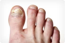
Certain nail conditions are often associated with ingrown toenails. For example, if you have had a toenail fungal infection or if you have lost a nail through trauma, you are at greater risk for developing an ingrown toenail.
Treatment
Sometimes initial treatment for ingrown toenails can be safely performed at home. However, home treatment is strongly discouraged if you suspect you have an infection, or if you have a medical condition that puts your feet at high risk for example; diabetes, nerve damage in the foot, or poor circulation.
Home care:
If you don’t have an infection or any of the above conditions, you can soak your foot in room—temperature water (add Epsoms salt if you wish), and gently massage the side of the nail fold to help reduce the inflammation.
Avoid attempting "bathroom surgery." Repeated cutting of the nail can cause the condition to worsen over time. If your symptoms fail to improve, it’s time to see a foot and ankle surgeon.
Physician care:
The foot and ankle surgeon will examine your toe and select the treatment best suited for you.
Treatment may include:
- Oral antibiotics. If an infection is present, an oral antibiotic may be prescribed.
- Surgery. A simple procedure, often performed in the office, is commonly needed to ease the pain and remove the offending nail. Surgery may involve numbing the toe and removing a corner of the nail, a larger portion of the nail, or the entire nail.
- Permanent removal. Various techniques may be used to destroy or remove the nail root. This treatment prevents the recurrence of an ingrown toenail. Your surgeon will determine the most appropriate procedure for you.
Following nail surgery, a light bandage will be applied, most people experience very little pain after surgery and may resume normal activity the next day. If your surgeon has prescribed an oral antibiotic, be sure to take all the medication, even if your symptoms have improved.
Preventing Ingrown Toenails
Many cases of ingrown toenails may be prevented by following these two important tips:
- Trim your nails properly. Cut your toenails in a fairly straight line, and don’t cut them too short. You should be able to get your fingernail under the sides and end of the nail.
- Avoid poorly-fitting shoes. Don't wear shoes that are short or tight in the toe box. Also avoid shoes that are loose, because they too cause pressure on the toes, especially when you run or walk briskly.
- Cutting a notch (a "V”) in the nail will reduce the tendency for the nail to curve downward the growth of the toenail. New cutting a “V" does not affect nail growth will continue to curve downward.
- Repeated trimming of the nail borders is a good way to’ treat ingrown toenails.
- Repeated nail trimming fails to correct future nail growth and can make the condition worse.
- Cotton placed under the nail will relieve the pain.
- Cotton placed under the nail can be harmful. It can easily harbour bacteria and encourage infection.
- You can buy effective ingrown toenail treatments at the drug store.
- Over-the-counter topical medications may mask the pain, but they fail to address the underlying problem.
What Is Hammertoe?
Hammertoe is a contracture or bending of one or both joints of the second, third, fourth, or fifth (little) toes. This abnormal bending can put pressure on the toe when wearing shoes, causing problems to develop. Common symptoms of hammertoes include:
- Pain of irritation of the affected toe when Wearing shoes.
- Corns (a build-up of slide) on the top, side, ‘or end of the toe, or between two toes. (Corns are caused by constant friction against the shoe. They may be soft or hard, depending upon their location.)
- Calluses (another type 'of skin build-up) on the bottom of the toe or on the ball of the foot.
Corns and calluses can be painful and make it difficult to find a comfortable shoe. But even without corms and calluses, hammertoes can cause pain because the joint itself may become dislocated.
Hammertoes usually start out as mild deformities and get progressively worse over time. In the earlier stages, hammertoes are flexible end the symptoms can often be managed with non-invasive measures. But if left untreated, hammertoes can become more rigid and will not respond to non—surgical treatment. Corns are more likely to:
Develop as time goes on—and corns never really go away, even after trimming. In more severe cases of hammertoe, open sores may form.
Because of the progressive nature of hammertoes, they should receive early attention. Hammertoes never get better without some kind of intervention.
- Normal Toes
- Hammertoes
What Causes Hammertoe?
The most common cause of hammertoe is a muscle/tendon imbalance, this imbalance, which leads to a bending of the toe, results from mechanical (structural) changes in the foot that occur over time in some people.
Hammertoes are often aggravated by shoes that don’t fit properly for example, shoes that crowd the toes. And in some cases, ill-fitting shoes can actually cause the contracture that defines hammertoe. For example, a hammertoe may develop it a toe is too long and is forced into a cramped position when a tight shoe is worn.
Occasionally, hammertoe is caused by some kind of trauma, such as a previously broken toe. In some people, hammertoes are inherited.
Treatment: Non-Surgical Approaches
There are a variety of treatment options for hammertoe. The treatment your pediatric foot and ankle surgeon selects will depend upon the severity of your hammertoe and other factors.
A number of non—surgical measures can be undertaken:
- Trimming cams and calluses. This should be done by a healthcare professional. Never attempt to, do this yourself, because you run the risk of cuts and infection. Your pediatric surgeon knows the proper way to trim corns to bring you the greatest benefit.
-
Padding cortisol calluses. Your podiatric surgeon can provide or prescribe pads designed to shield corn's from irritation. If you want to try over-the-counter pads, avoid the medicated types. Medicated pads are generally not recommended because they may contain a small amount of acid that can be harmful. Consult your podiatric surgeon about this option.
-
Changes in shoe wear. Avoid shoes with pointed toes, shoes that are too short, or shoes with high heels—conditions that can force your toe against the front of the shoe. Instead, choose comfortable shoes with a sleep, roomy toe box and heels no higher than two inches.
-
Orthotic devices. A Custom orthotic device placed in your shoe may help control in the muscle/tendon imbalance.
-
Injection therapy. Corticosteroid injections are sometimes used to ease pain and inflammation caused by hammer toe.
-
Medications. Nonsteroidal antiinflammatory drugs (NSAIDS), such as ibuprofen, are; often prescribed to reduce pain and inflammation.
-
Splinting/strapping. Splints or small straps may be applied by the podiatric surgeon to realign the bent toe.
When Is Surgery Needed?
In some cases, usually when the hammertoe has become more rigid, surgery is needed to relieve the pain and discomfort caused by the deformity. Your podiatric surgeon will discuss the options and select a plan tailored to your needs. Among other concerns, he-or she will take into consideration the type of shoes you want to wear, the number of toes involved, your activity level, your age, and the severity of the hammertoe.
The most Common surgical procedure performed to correct a hammertoe is called arthroplasty. In this procedure, the surgeon removes a small section of the bone from the affected joint.
Another surgical option is arthrodesis, which is usually reserved for more rigid toes or severe cases, such as when there are multiple joints or toes involved.
Arthrodesis is a procedure that involves a fusing of a small joint in the toe to straighten it. A pin or other small fixation device is typically used to hold the toe in position while the bones are healing.
It is possible that a patient may require other procedures, as well especially when the hammertoe condition is severe. Some of these procedures include skin wedging (the removal-of wedges of skin), tendon/muscle rebalancing or lengthening, small tendon transfers, or relocation of surrounding joints.
Often patients with hammertoe have bunions or other foot deformities corrected at the same time. The length of the recovery period will vary, depending on the procedure or procedures performed.
Foot Health
- Achilles Tendonitis
- Ankle Instability
- Ankle Sprains
- Arthritis Foot & Ankle Care
- Athletes Foot
- Bunions
- Calluses
- Corns
- Crush Injuries
- Diabetic Foot Care
- Flat Feet
- Fractures
- Fungus Toenails (Laser treatment)
- Geriatric Foot Care
- Hammertoes
- Heel Pain
- Ingrown Toenails
- Injuries
- Neuromas
- Orthotics
- Plantar Fasciitis
- Sports Medicine
- Surgery
- Warts
Communities we serve
- Wildflower Court
- Ketchikan Pioneer Home
- Sitka Pioneer Home
- Petersburg Pioneer Home
- Juneau/Auke Bay/Douglas
- Ketchikan Clinic
- Metlakatla
- Haines
- Skagway
- Sitka
- Wrangell
- Angoon
- Petersburg
- Gustavus
- Yakutat
Lisfranc Injuries
The Lisfranc joint
The Lisfranc joint is the point at which the metatarsal bones (long bones that lead up to the toes) and the tarsal bones (bones in the arch) connect. The Lisfranc ligament is a tough band of tissue that joins two of these bones. It is important for maintaining proper alignment and strength of this joint.
How do Lisfranc Injuries Occur?
Injuries to the Lisfranc joint most commonly occur in automobile accident victims, military personnel, runners, horseback riders, football players, and participants of other contact sports. Lisfranc injuries occur as a result of direct or indirect forces to the foot. A direct force often involves something heavy falling on the foot. Indirect force commonly involves twisting the foot. This can happen, for example, when the foot catches on a stirrup while falling from a horse.
Types of Lisfranc Injuries
There are three types of Lisfranc injuries, which sometimes occur together:
- Sprains. The Lisfranc ligament, as well as other ligaments on the bottom of the midfoot, are stronger than the ligaments on the top of the midfoot. Therefore, when they are weakened through a sprain (a stretching of the ligament), patients experience instability of the joint in the middle of the foot.
- Fractures. A break in a bone in the Lisfranc joint can be either an avulsion fracture (a small piece of bone is pulled off) or a break through the bone or bones of the midfoot.
- Dislocations. The bones of the Lisfranc joint may be forced from their normal positions.
Signs and Symptoms
The signs and symptoms of a Lisfranc injury may include:
- Swelling of the foot
- Pain throughout the midfoot when standing or when pressure is applied
- Inability to bear weight (in severe injuries)
- Bruising or blistering on the arch this is an important sign- of a Lisfranc injury. Bruising may also occur on the top of the foot.
- Abnormal widening of the foot.
Diagnosis
Lisfranc injuries are sometimes mistaken for ankle sprains, making the diagnostic process very important. To arrive at a diagnosis, the foot and ankle surgeon will ask how the injury occurred. The surgeon will examine the foot and determine the severity of the injury.
X-rays and other imaging studies such as a CT or MRI may be necessary to fully evaluate the extent of the injury. The surgeon may also perform an additional examination While the patient is under anesthesia to further evaluate a fracture or weakening of the joint and surrounding bones.
Treatment
Anyone who has symptoms of a Lisfranc injury should see a foot and ankle surgeon right away. If unable to do so immediately, it is important to stay off the injured foot, keep it elevated (at or slightly above hip level), and apply a bag of ice wrapped in a thin towel to the area every 20 minutes of each waking hour. These steps will help keep the swelling and pain under control.
Treatment by the foot and ankle surgeon may include one or more of the following, depending on the type and severity of the Lisfranc injury:
- Immobilization. Sometimes the foot is placed in a cast to keep it immobile, and crutches are used to avoid putting weight on the injured foot.
- Oral medications. Nonsteroidal anti-inflammatory medications (NSAIDs), such as ibuprofen, help reduce the pain and inflammation.
- Ice and elevation. Swelling is reduced by icing the affected area and keeping the foot elevated, as described above.
- Physical therapy. After the swelling and pain have subsided, physical therapy may be prescribed.
- Surgery. Certain types of Lisfranc injuries require surgery. The foot and ankle surgeon will determine the type of procedure that is best suited to the individual patient. Some injuries of this type may require emergency surgery.
Complications of Lisfranc Injuries
Complications can and often do arise following Lisfranc injuries. A possible early complication following the injury is compartment syndrome, in which pressure builds. up within the tissues of the foot, requiring immediate surgery to prevent tissue damage. A build-up of pressure could damage the nerves, blood vessels, and muscles in the foot.
Arthritis and problems with foot alignment may develop. In most cases, arthritis develops several months or longer following a Lisfranc injury, requiring additional treatment.
Malignant Melanoma of the Foot
What is Malignant Melanoma?
Melanoma is a cancer that begins in the cells of the skin that produce pigmentation (coloration). It is also called malignant melanoma because it spreads to other areas of the body as it grows beneath the surface of the skin. Unlike many other types of cancer, melanoma strikes people of all age groups, even the young.
Melanoma in the Foot
Melanoma that occurs in the foot or ankle often goes unnoticed during its earliest stage, when it would be more easily treated. By the time melanoma of the foot or ankle is diagnosed, it frequently has progressed to an advanced stage, accounting for a higher mortality rate. This makes it extremely important to follow prevention and early detection measures involving the feet as well as other parts of the body.
Causes
Most cases of melanoma are caused by too much exposure to ultraviolet (UV) rays from the sun or tanning beds. This exposure can include intense UV radiation obtained during short periods, or lower amounts of radiation obtained over longer periods.
Anyone can get melanoma, but some factors put a person at greater risk for developing this type of cancer. These include:
- Fair skin; skin that freckles; blond or red hair
- Blistering sunburns before the age of 18
- Numerous moles, especially if they appeared at a young age
What Should You Look For?
Melanoma can occur anywhere on the skin, even in areas of the body not exposed to the sun. Melanoma usually looks like a spot on the skin that is predominantly brown, black, or blue--- although in some cases it can be mostly red or even white. However, not all areas of discoloration on the skin are melanoma.
There are four signs---known as the ABCDs of melanoma---to look for when self-inspecting moles and other spots on the body:
Asymmetry
Melanoma is usually asymmetric, which means one half is different in shape from the other half.
Border
Border irregularity often indicates melanoma. The border---or edge---is typically ragged, notched, or blurred.
Color
Melanoma is typically a mix of colors or hues, rather than a single, solid color.
Diameter
Melanoma grows in diameter, whereas moles remain small. A spot that is larger than 5 millimeters (the size of a pencil eraser) is a cause for concern.
If any of these signs are present on the foot, it is important to see a foot and ankle surgeon right away. It is also essential to see a surgeon if there is discoloration of any size underneath a toenail (unless the discoloration was caused by trauma, such as stabbing a toe or having something fall on it).
Diagnosis
To diagnose melanoma, the foot and ankle surgeon will ask the patient a few questions. For example: Is the spot old or new? Have you noticed any changes in Size or color? If so, how rapidly has this change occurred?
The surgeon will also examine the spot to determine whether a biopsy is necessary. If a biopsy is performed and it reveals melanoma, the surgeon will discuss a treatment plan.
Prevention and Early Detection
Everyone should practice strategies that can help prevent melanoma---or at least aid in early detection, so that early treatment can be undertaken
Precautions to avoid getting melanoma of the foot and ankle, as well as general precautions, include:
- Wear water shoes or shoes and socks---flip flops do not provide protection!
- Use adequate sunscreen in areas that are unprotected by clothing or shoes. Be sure to apply sunscreen on the soles as well as the tops of feet.
- Inspect all areas of the feet daily--- including the soles, underneath toenails, and between the toes.
- If you wear nail polish, remove it occasionally so that you can inspect the skin underneath the toenails
- Avoid UV radiation during the sun’s peak hours (10 am. to 4 p.m.), beginning at birth. While sun exposure is harmful at any age, it is especially damaging to children and adolescents.
- Wear sunglasses that block 100% of all UV rays---both UVA and UVB.
- Wear a wide-brimmed hat.
Remember: Early detection is crucial with malignant melanoma. If you see any of the ABCD signs---or if you have discoloration beneath a toenail that is unrelated to trauma--- be sure to visit a foot and ankle surgeon as soon as possible.
Posterior Tibial Tendon Dysfunction
What Is PTTD?
Posterior tibial tendon dysfunction (PTTD) is an inflammation and / or overstretching of the posterior tibial tendon in the foot. An important function of the posterior tibial tendon is-to help support the arch. But in PTTD, the tendons ability to perform that job is impaired, often resulting in a flattening of the foot.
The posterior tibial tendon is a fibrous cord that extends from a muscle in the leg, It descends the leg and runs along the inside of the ankle, down the side of the foot, and into the arch. This tendon serves as one of the major supporting structures of the foot and helps the foot to function while walking.
PTTD is often called “adult- acquired flatfoot" because it is the most common type of flatfoot developed during adulthood. Although this condition typically occurs in only one foot, some people may develop it in both feet. PTTD is usually progressive, which means it will keep getting worse --- especially if it isn’t treated early.
Symptoms of PTTD
The symptoms of PTTD may include pain, swelling, a flattening of the arch, and an inward rolling of' the ankle. As the condition progresses, the symptoms will change.
For example:
- When PTTD initially develops, typically there is pain on the inside of the foot and ankle (along the course of the tendon). In addition, the area may be red, warm, and swollen.
- Later, as the arch begins to flatten, there may still be a pain on the inside of the foot and ankle. But at this point, the foot and toes begin to turn outward and the ankle rolls inward.
- As PTTD becomes more advanced, the arch flattens even more and the pain often shifts to the outside of the foot, below the ankle. The tendon has deteriorated considerably and arthritis often develops in the foot. In more severe cases, arthritis may also develop in 'the ankle.
What Causes PTTD?
Overuse of the posterior tibial tendon is frequently the cause of PTTD. In .fact, the symptoms usually occur after activities that involve the tendon, such as running, walking, hiking, or climbing stairs.
Treatment: Non-surgical Approaches
Because of the progressive nature of PTTD, it’s best to see your foot and ankle surgeon as soon as possible. If treated early enough, your symptoms may resolve without the need for surgery and progression of your condition can be arrested. In contrast, untreated PTTD could leave you with an extremely flat foot, painful arthritis in the foot and ankle, and increasing limitations on walking, running, or other activities.
In many cases of PTTD treatment can begin with nonsurgical approaches that may include:
- Orthotic devices or bracing. To give your arch the support it needs, your foot and ankle surgeon may provide you with an ankle stirrup brace or a custom orthotic device that fits into the shoe.
- Immobilization. Sometimes a short-leg cast or boot is worn to immobilize the foot and allow the tendon to-heal, or you may need to completely avoid all weight- bearing for a while.
What Are the Peroneal Tendons?
A tendon is a band of tissue that connects a muscle to a bone. In the foot, there are two peroneal tendons. They run side-by-side behind the outer ankle bone. One peroneal tendon attaches to the other part of the midfoot, while the other tendon runs under the foot and attaches near the inside of the areh. The main function of the peroneal tendons is to stabilize the foot and ankle and protect them from sprains.
Types of Peroneal Tendon Injuries
Peroneal tendon injuries may be acute (occurring suddenly) or chronic (developing over a period of time), They most commonly occur in individuals who participate in sports that involve repetitive ankle motion In addition, people with higher arches are at risk for developing peroneal tendon injuries. Basic types of peroneal tendon injuries are tendonitis, tears, and subluxation.
Tendonitis is an inflammation of one or both tendons. The inflammation is caused by activities involving repetitive use of the tendon, overuse of the tendon, or trauma (such as an ankle sprain).
Symptoms of tendonitis include:
- Pain
- Swelling
- Warmth to the touch
Acute tears are caused by repetitive activity or trauma. Immediate symptoms of acute tears include:
- Pain
- Swelling
- Weakness or instability of the foot and ankle
As time goes on, these tears may lead to a change in the shape of the foot, in which the arch may become higher.
Degenerative teats (tendinosis) are usually due to overuse and occur over long periods of time---often years. In degenerative tears, the tendon is like taffy that has been overstretched until it becomes thin and eventually trays. Having high arches also puts you at risk for developing a degenerative tear. The signs and symptoms of degenerative tears may include:
- Sporadic pain (occurring from time to time) on the outside of the ankle
- Weakness or instability in the ankle
- An increase in the height of the arch
Subluxation - one or both tendons have slipped out of their normal position. In some cases, subluxation is due to a condition in which a person is born with a variation in the shape of the bone or muscle. In other cases, subluxation occurs following trauma, such as an ankle sprain. Damage or injury to the tissues that stabilize the tendons (retinaculum) can lead to chronic tendon subluxation. The symptoms of subluxation may include:
- A snapping feeling of the tendon around the ankle bone
- Sporadic pain behind the outside ankle bone
- Ankle instability or weakness
Early treatment of a subluxation is critical, since a tendon that continues to sublux (move out of position) is more likely to tear or rupture. Therefore, if you feel the characteristic snapping, see a foot and ankle surgeon immediately.
Diagnosis
Because peroneal tendon injuries are sometimes misdiagnosed and may worsen without proper treatment, prompt evaluation by a foot and ankle surgeon is advised; To diagnose a peroneal tendon injury, the surgeon will examine the foot and look for pain, instability, swelling, warmth, and weakness on the outer side of the ankle. In addition, imaging studies such as an MRI or ultrasound may be needed to fully evaluate the injury. An ankle sprain may sometimes accompany a peroneal tendon injury. The surgeon is trained to look for signs of this and other related injuries. Proper diagnosis; is important because prolonged discomfort after a simple sprain may be a sign of additional problems.
Treatment
Treatment depends on the type of peroneal tendon injury. Options include:
- Immobilization. A cast or splint may be used to keep the foot and ankle from moving and allow the injury to heal.
- Medications. Oral or injected anti-inflammatory drugs may help relieve the pain and inflammation.
- Physical therapy. Ice, heat, or ultrasound therapy may be used to reduce swelling and pain As symptoms improve, exercises can be added to strengthen the muscles and improve range of motion and balance.
- Bracing. The surgeon may provide a brace to use for a short While or during activities requiring repetitive ankle motion. Bracing may also be an option when a patient is not a candidate for surgery.
- Surgery. In some cases, surgery may be needed to repair the tendon or tendons and perhaps the supporting structures of the foot. The foot and ankle surgeon will determine the most appropriate procedure for the patient’s condition and lifestyle. After surgery, physical therapy is an important part of rehabilitation.
What Is Osteoarthritis?
Osteoarthritis is a condition characterized by the breakdown and eventual loss of cartilage in one or more joints, Cartilage---the connective tissue found at the end of the bones in the joints---protects and cushions the bones during movement. When cartilage deteriorates or is lost, symptoms develop that can restrict one's ability to easily perform daily activities.
Osteoarthritis is also known as degenerative arthritis, reflecting its nature to develop as part of the aging process. As the most common form of arthritis, osteoarthritis affects millions of Americans. Many people refer to osteoarthritis simply as arthritis, even though there are more than 100 different types of arthritis.
Osteoarthritis appears at various joints throughout the body; including the hands, feet, spine, hips, and knees. In the foot, the disease most frequently occurs in the big toe, although it is also often found in the match and ankle.
Signs and Symptoms
People with osteoarthritis in the foot or ankle experience, in varying degrees, one or more of the following:
- Pain and stiffness in the joint
- Swelling in or near the joint
- Difficulty walking or bending the joint
Some patients with osteoarthritis also develop a bone spur (a bony protrusion) at the affected joint. Shoe pressure may cause pain at the site of a bone spur, and in some cases blisters or calluses may form over its surface. Bone spurs can also limit the movement of the joint.
Causes
Osteoarthritis is considered a "wear and tear” disease because the cartilage in the joint wears down with repeated stress and use over time. As the cartilage deteriorates and gets thinner, the bones lose their protective covering and eventually may rub together, causing pain and inflammation of the joint.
An injury may also lead to osteoarthritis, although it may take months 'or years after the injury for the condition to develop. For example, osteoarthritis in the big toe is often caused by kicking or jamming the toe, or by dropping something on the toe. Osteoarthritis in the midfoot is often caused by dropping something on it, or by a sprain or fracture, In the ankle, osteoarthritis is usually caused by a fracture and occasionally by a severe sprain.
Sometimes osteoarthritis develops as a result of abnm'mal foot mechanics. People who have flat feet or high arches are at increased risk for developing osteoarthritis in the foot.
A flat foot causes less stability in the ligaments (bands of tissue that connect bones), resulting in excessive strain on the joints, which can cause arthritis. A high arch is rigid and lacks mobility, causing a jamming of joints that creates an increased risk of arthritis.
Diagnosis
In diagnosing osteoarthritis, the foot and ankle surgeon will examine the foot thoroughly, looking for swelling in the joint, limited mobility, and pain with movement. In some cases, deformity and/ or enlargement (spur) of the joint may be noted.
What Is an Ankle Sprain?
An ankle sprain is an injury to one or more ligaments in the ankle, usually on the outside of the ankle. Ligaments are bands of tissue---like rubber bands---that connect one bone to another and bind the joints together: In the ankle joint, ligaments provide stability by limiting side-to-side movement. Some ankle sprains are much worse than others. The severity of an ankle sprain depends on whether the ligament is stretched, partially torn, or completely torn, as well as on the number of ligaments involved. Ankle sprains are not the same as strains, which affect muscles rather than ligaments.
What Causes a Sprained Ankle?
Sprained ankles often result from a fall, a sudden twist, or a blow that forces the ankle joint out of its normal position. Ankle sprains commonly occur while participating in sports, wearing inappropriate shoes, or walking or running on an uneven surface.
Sometimes ankle sprains occur because of weak ankles, a condition that some people are born With. Previous ankle or foot injuries can also weaken the ankle and lead to sprains.
Signs and Symptoms
The signs and symptoms of ankle sprains may include:
- Pain or soreness
- Swelling
- Bruising
- Difficulty walking
- Stiffness in the joint
These symptoms may vary in intensity depending on the severity of the sprain. Sometimes pain and swelling are absent in people With previous ankle sprains---instead, they may simply feel the ankle is wobbly and unsteady when they walk. Even if you don't have pain or swelling with a sprained ankle, treatment is crucial. Any ankle sprain---whether it’s your first or your fifth requires prompt medical attention.
If you think you’ve sprained your ankle, contact your foot and ankle surgeon for an appointment as soon as possible. In the meantime, immediately begin using the "RICE” method (Rest, Ice, Compression, and Elevation) to help reduce swelling, pain, and further injury.
Why Prompt Medical Attention Is Needed
There are four key reasons why an ankle sprain should be promptly evaluated and treated by a foot and ankle surgeon:
- First, an untreated ankle sprain may lead to chronic ankle instability, a condition marked by persistent discomfort and a "giving way” of the ankle. You may also develop weakness in the leg.
- Second, you may have suffered a more severe ankle injury along with the sprain, This might include a serious bone fracture that could lead to troubling complications if it goes untreated.
- Third, an ankle sprain may be accompanied by a foot injury that causes discomfort but has gone unnoticed thus far.
- Fourth, rehabilitation of a sprained ankle needs to begin
What Is an Ankle Fracture?
A fracture is a partial or complete break in a bone. In the ankle, fractures cart range from the less serious avulsion injuries (small pieces of bone that have been pulled off) to severe shattering-type breaks of the tibia, fibula, or both.
Ankle fractures are common injuries that are most often caused by the ankle rolling inward or outward. Many peopleE mistake an ankle fracture for an ankle sprain, but they are quite different and therefore require an accurate and early diagnosis. They sometimes occur simultaneously.
Signs and Symptoms
An ankle fracture is accompanied by one or all of these signs and symptoms:
- Pain at the site of the fracture, Which in some cases can extend from the foot to the knee
- Significant swelling, which may occur along the length of the leg of may be more localized
- Blisters may occur over the fracture site. These should be promptly treated by your surgeon
- Bruising, which develops soon after the injury
- Inability to walk---however, it is possible to walk With less severe breaks, so never rely on walking as a test of whether a bone has been fractured
- Change in the appearance of the ankle so that it differs from the other ankle
- Bone protruding through the skin---a sign that immediate care is needed! Fractures that pierce the skin require immediate attention because they can lead to severe infection and prolonged recovery.
What to Do
Following an ankle injury it is important to have the ankle evaluated by a foot and ankle surgeon for proper diagnosis and treatment. If you are unable to do so right away, go to the emergency room and then follow up with a foot and ankle surgeon as soon as possible for a more thorough assessment.
Until you are able to be examined by a doctor; the “RICE." principle should be followed. This involves:
- Rest. It is crucial to stay off the injured foot, since walking can cause further damage.
- Ice. To reduce swelling and pain, apply a bag of ice over a thin towel to the affected area for 20 minutes of each waking hour. Do not put ice directly against the skin
- Compression. Wrap the ankle in an elastic bandage or wear a compression stocking to prevent further swelling.
- Elevation. Keep the foot elevated to reduce the swelling. It should be even with or slightly above the hip level.
Diagnosis
The foot and ankle surgeon will examine the affected limb, touching specific areas to evaluate the injury. In addition, the surgeon may order x-rays and other imaging studies, as necessary.
Treatment
Treatment of ankle fractures depends upon the type and severity of the injury. Options include:
- Immobilization. Certain fractures are treated by protecting and restricting the ankle and foot in a cast or splint. This allows the bone to heal.
- Prescription medications. To help relieve the pain, the surgeon may prescribe pain medications or anti-inflammatory drugs.
- Surgery. For some injuries, surgery is needed to repair the fracture and other related injuries, if present. The foot and ankle surgeon will select the procedure that is appropriate for your injury.
Follow-Up Care
It is important to follow your surgeon’s instructions after treatment. Failure to do so can lead to infection, deformity, arthritis, and chronic pain.
Other Conditions We Can Help Treat
Apart from the exhaustive list of foot injuries and conditions we have discussed above, you can also come to our clinic for other foot-related concerns. We provide expert medical advice and assistance for the following:
- Foot concerns related to diabetic conditions
- Foot neuroma
- Foot odor
- Foot orthoses
- Fungal nails or fungus nails
- Ganglion
- Gout
- Injuries and other concerns regarding children’s feet
- Planar fasciitis
- Sweaty feet
Southeast Foot & Ankle Center
Anh T. Lam, D.P.M.
7691 Glacier Hwy.
Juneau, AK 99801
Phone Number: 907-789-5518
Fax Number: 907-523-6991